Osteoblast niches
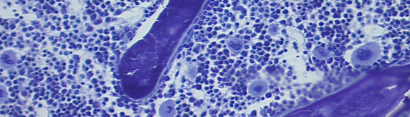
Osteoblast niches
The mechanisms involved in the defective bone formation with age are largely unknown (Kassem & Marie, 2009). Mesenchymal stromal cell (MSC) adhesion, proliferation and differentiation are highly dependent on cell-cell interactions and signaling mechanisms. Recent studies highlighted the importance of niches as physiologial entities that control stem cell differentiation in the adult bone marrow (Wu et al., 2009). However, the mechanisms that regulate the formation and activity of MSC in their niche during aging and disease are unknown (Adams et al., 2007). This project aims to investigate MSC/osteoblast interactions during bone aging and disease. We previously demonstrated that N-cadherin interacts with Wnt signaling to control osteoblastogenesis and bone accrual (Haÿ et al., 2009a,b; Marie, 2009). In this project, we will test the hypothesis that N-cadherin expressed in osteoblasts controls the MSC pool and osteogenic differentiation during aging, as well as in a pathological context such as cancer. Our first objective is to determine the implication of N-cadherin expressed by osteoblasts on MSC pool within what we call the « osteogenic niche », where MSC and mature osteoblasts interact in the bone marrow. Using our N-cadherin transgenic mice, we will determine whether N-cadherin overexpression in osteoblasts impacts MSC cell adhesion, growth, differentiation and survival in the osteogenic niche in mice, in relation to the age-related bone loss. To this goal, primary bone marrow stromal cells isolated from syngenic mice will be injected in long bones of WT or N-cadherin Tg neonatal and adult mice. After 18 weeks, MSC cell growth, differentiation and survival will be determined by histochemical analysis. In parallel, bone formation and bone mass will be determined by histomorphometric analysis after double fluorochrome labeling. The results will bring novel knowledge on the implication of N-cadherin in the local mechanisms controlling the osteogenic differentiation of MSC in relation to the age-related decrease in bone formation and bone loss. The second objective is to investigate the role of N-cadherin expressed by osteoblasts in bone cancer cell proliferation and invasion. To this goal, we will use murine (K7M2) invasive osteosarcoma cells, and analyse how these cancer cells behave when they are in contact with murine osteoblasts with low or high N-cadherin expression levels in vitro. We will determine the involvement of N-cadherin, using a neutralizing N-cadherin antibody. To determine the impact of N-cadherin expression on cancer cell progression in vivo, we will transplant murine K7M2-GFP cells in young WT or Tg mice into long bones, and K7M2-GFP cells growth and survival will be determined after 6 weeks by FACS analysis and histochemical analysis. Bone tumor growth and invasion will be determined by histological analysis and the occurrence of lung micrometastasis will be analysed. This will give essential information on the role of endogenous and exogenous (Tg) N-cadherin expressed by osteoblasts on cancer cell adherence, growth and dissemination. Overall, the results may allow us to develop therapeutic strategies targeting N-cadherin for sustaining MSC osteogenic differentiation as well as restraining cancer cell progression.
Références bibliographiques
1. Adams GB,et al Nat Biotechnol. 2007 Feb;25(2):238-43. Epub 2007 Jan 21. Erratum in: Nat Biotechnol. 2007
2. Aug;25(8):944. Nat Biotechnol. 2008 Feb;26(2):241.
3. Chapurlat RD, Orcel P, 2008, Best Pract Res Clin Rheumatol. 22(1):55-69.
4. d’Alésio A et al. Hum Mol Genet. 200, 14:3539-48.
5. Delgado-Calle J et al, 2012, Epigenetics. 7:83-91.
6. Delgado-Calle J, et al, 2012, J Bone Miner Res. 27:926-37.
7. Fang Y,et al, 2005, Am. J. Hum. Genet. 77: 807-823.
8. Geoffroy V et al, 2002, Mol. Cell. Biol. 22 (17): 6222–6233.
9. Haÿ E et al, 2009 , Mol Cell Biol. 29, 4: 953-964.
10. Haÿ E et al, 2009, PloSOne 4(12):e8284.
11. Jehan F et al, J. Clin. Endoc. Metabol. 93: 4672–4682.
12. Kassem M, Marie PJ, 2011, Aging Cell, 1474-9726.
13. Kim MS et al, 2009, Nature, 461(7266):1007-12.
14. Kocemba KA et al, PLoS One. 2012;7(2):e30359. Epub 2012 Feb 17
15. Locker M et al, 2006, Cell Signal. 2006 May;18(5):628-39.
16. Marie PJ.2009. IBMS BoneKEy. 6(4):150-6.
17. Merciris Det al, 2007, Am J Pathol, 170(5):1676-1685
18. Ogita M et al, 2008, Endocrinology, 149(11):5713-23
19. Samee N et al, 2008, Am J pathol 173(3):773-80.
20. Samee N et al, 2009, J Cell Biochem, 107(5):865-72
21. Wu JY et al, 2009, J Bone Miner Res. 24(5):759-64.
22. Wu JY et al, 2011, J Clin Invest. 121(9):3492-504