Histology
Histology
CONTACT
Mylène ZARKA
mylene.zarka@inserm.fr
Histomorphometry of hard tissue
The inclusion of hard tissues requires a technical specificity. Non decalcified samples (bones, vessel, meniscus and mineralizing tissues) are included in methacrylate‘s resin. Human biopsies are analyzed by histomorphometry.
In our lab, the histology of mineralizing tissues is in particular used for animal models using parameters of bone microarchitecture and activity. In our mouse models , we perform paraffin and frozen embedding of both bone and joints and then perform immunochemistry and immunofluorescent staining.
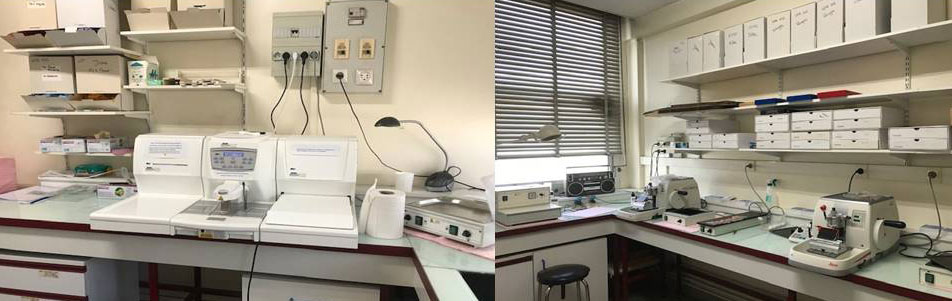
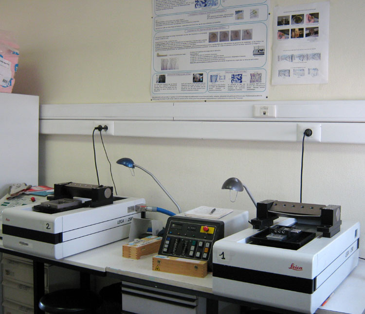
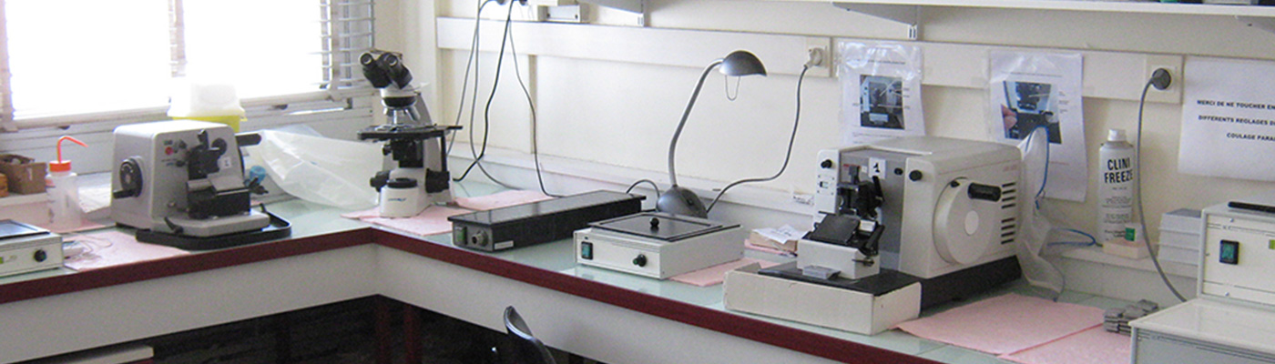
LEICA SM2500 microtomes for resine sectionning Paraffin and section inclusion sector
Quantification-imaging
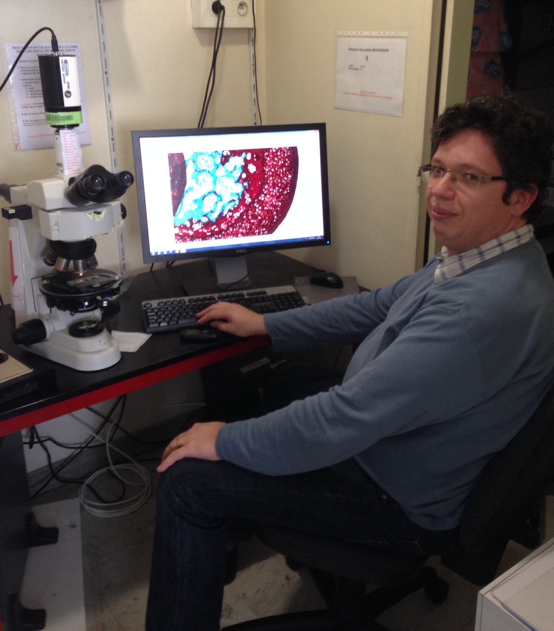
Bone histomorphometry is a technique which allows to quantify different parameters of bone microarchitecture and remodeling in murine and human samples. It allows the analysis of qualitative and quantitative parameters on histological sections of non decalcified bone which is necessary to explore bone cells and tissue « in vivo ».
Our histological center has 4 microscopes among which 2 are coupled to a camera and a computer with the programs BONOLAB and HISTOLAB (MICROVISION®).It is used for the quantitative analysis of both bone and joints histomorphometry.
Specific bone and joint stainings
Non decalcified tissue sections embedded in metacrylate’s resine :
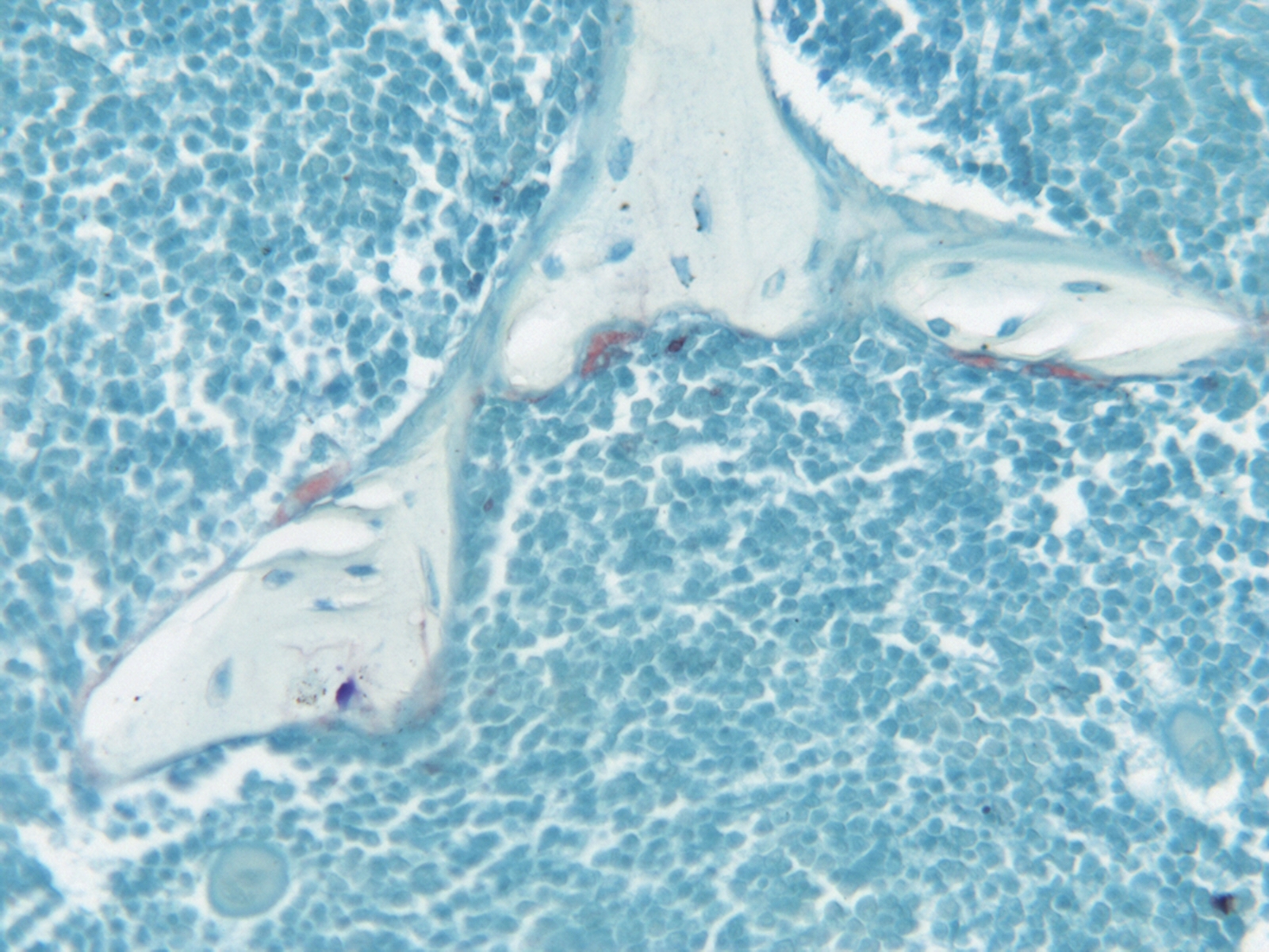
Tartrate resistant acid phosphatases positive cells (osteoclast activity).
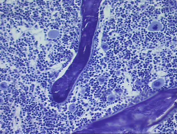
Toluidin blue staining showing the bone cells and the bone structure
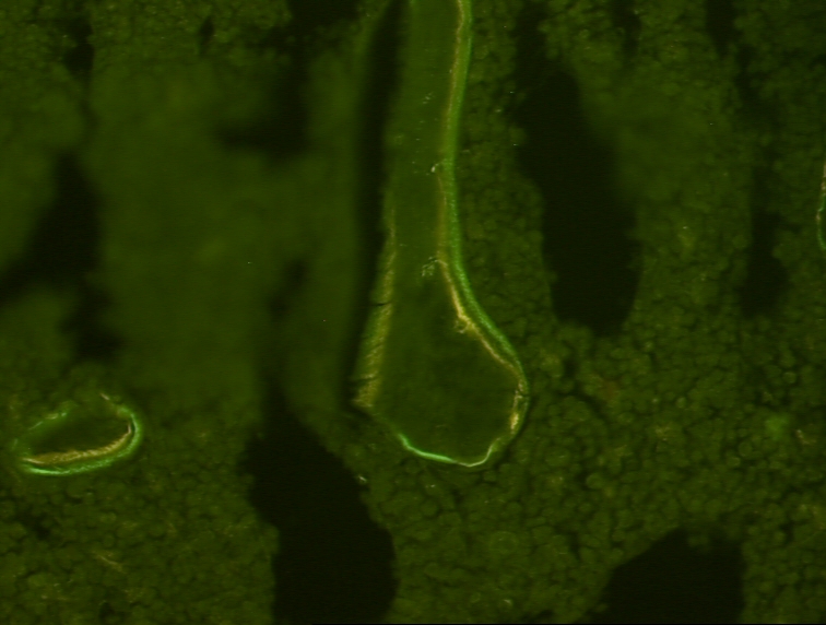
Tetracycline and calceine injections allow the evaluation of the dynamic parameters of bone.
Decalcified tissue sections embedded in paraffin :
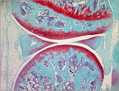
Safranine-O staining of murine joint (Proteoglycans)
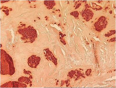
Red alizarin staining of human meniscus