Imaging platform
Plateforme collaborative SFR IMOSAR : www.imosar.cnrs.fr
Imaging platform
We are focusing our research on the bone and joint field, with a strong relationship with clinical research.
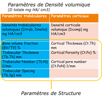
Our imaging methods help us to develop biomarkers. These markers are quantitative tools with the objective to follow structural variations in pathological and normal bone and joint tissues. One of the medical imaging apparatus used in our laboratory is the scanner (CT-scanner). This tool is using X-rays that are electromagnetic radiations composed with high energy photons being used in medical research for diagnostic purposes.
CONTACT
Agnès OSTERTAG
agnes.ostertag@inserm.fr
In Human
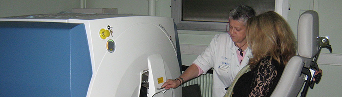
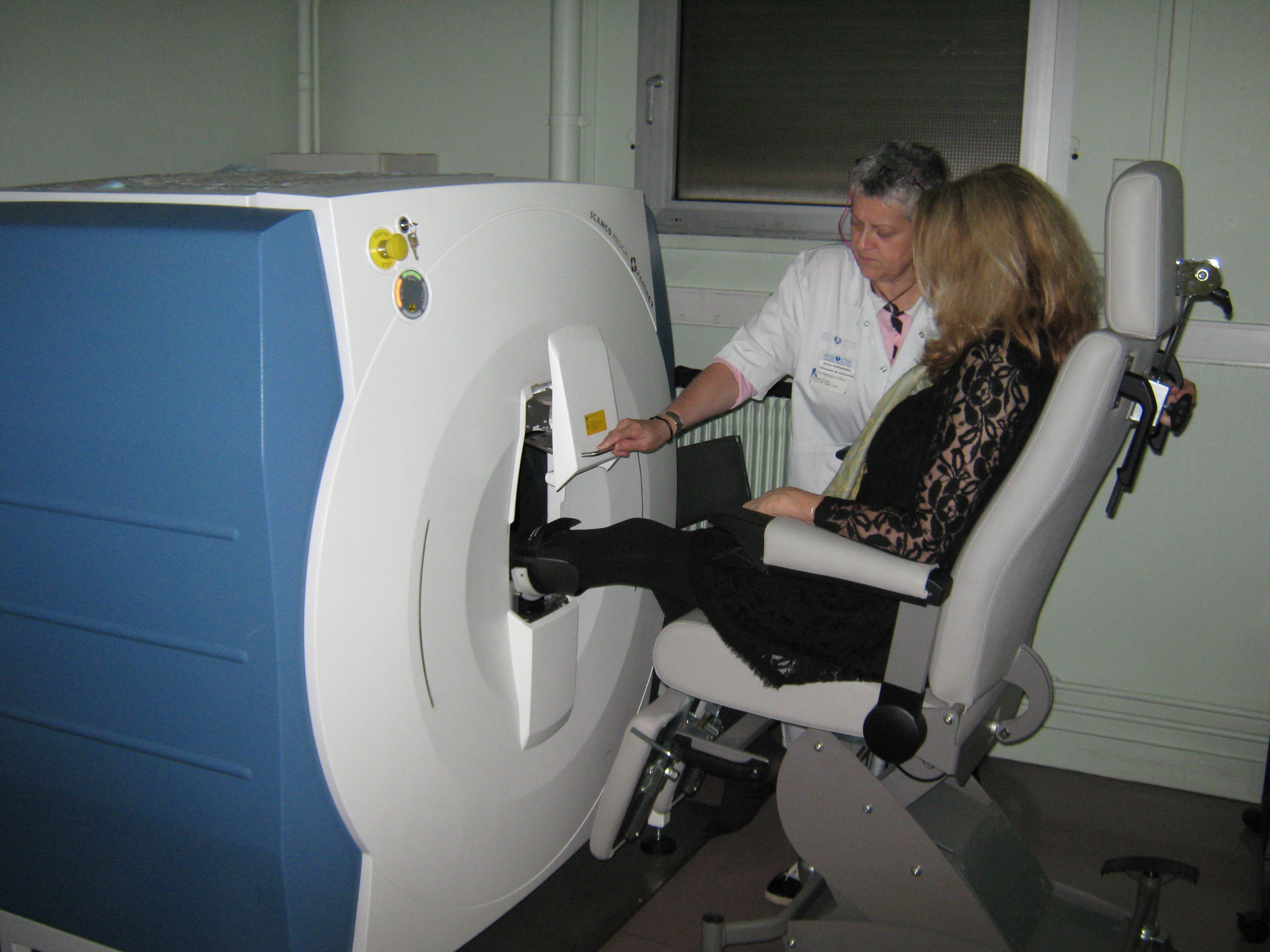
XTREMCT SCANCO
Peripheral Quantitative Tomography using X-rays to study the micro-architectural bone: high-resolution 3-D imaging (80µm3) of human bones (Radius and Tibia) for the diagnostic and the follow-up of osteoporosis disease.
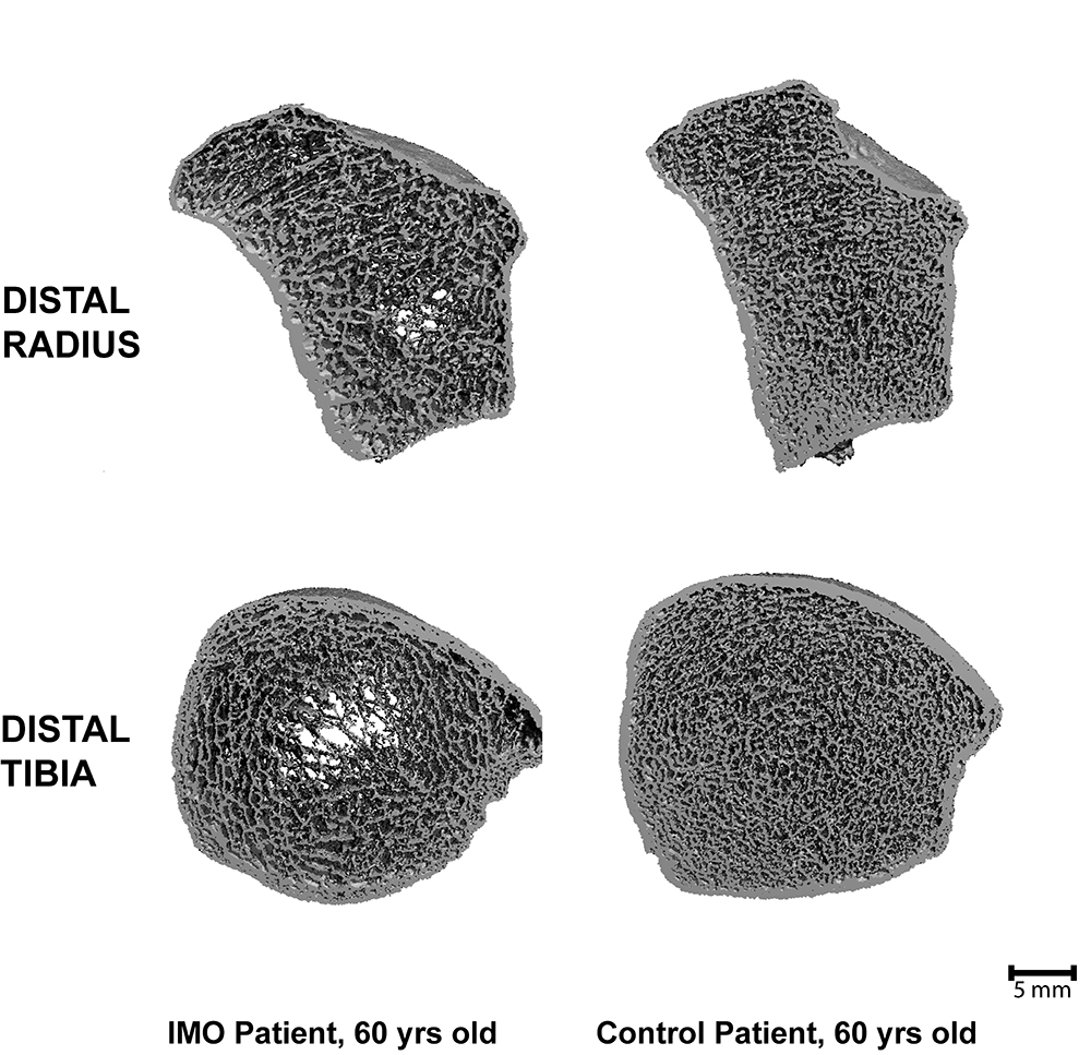
Image above : On the left, patient with an idiopathic osteoporosis (decrease number and thinning of trabeculae), on the right, healthy patient (numerous and well connected trabeculae)
In Animal
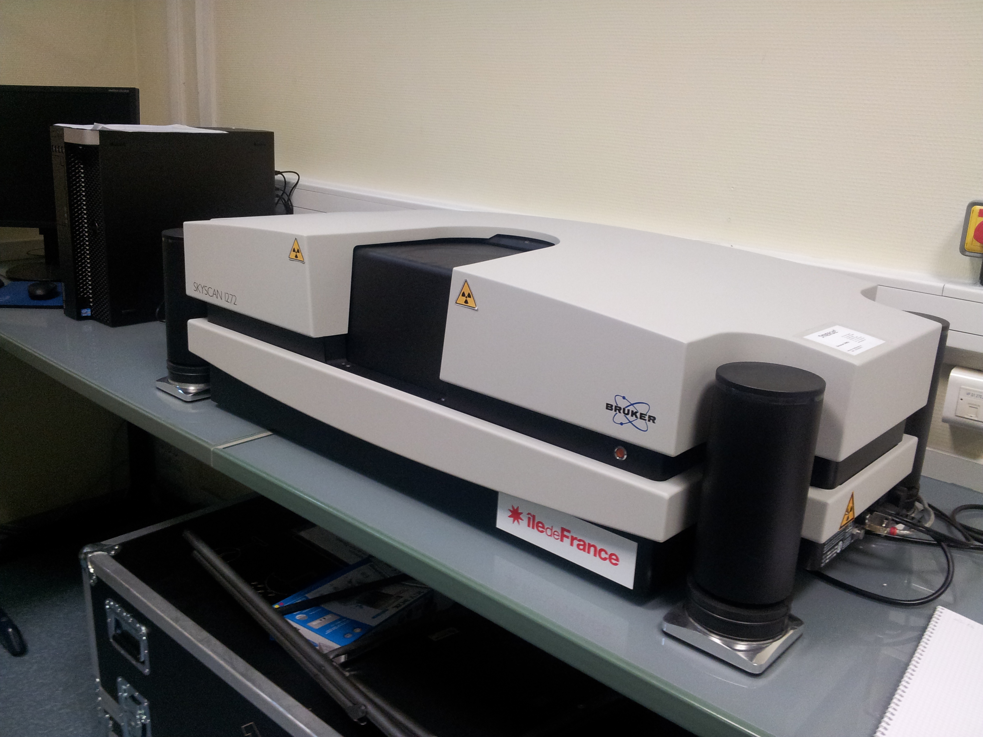
MICRO-SCANNER IN VITRO BRUKER SKYSCAN 1272
Descriptive : High-resolution 3-D imaging (5 to 30 µm3) with X-rays in animal or human tissues with or without integrated biological material.
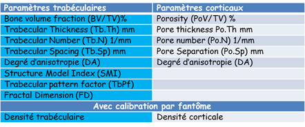
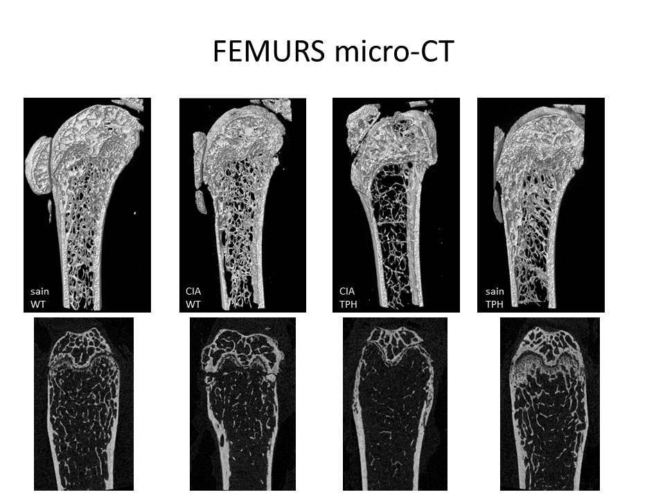
Study of vertebral or femoral bone microarchitecture
Femurs of mices : Up level = 3D images, down level = 2D slices
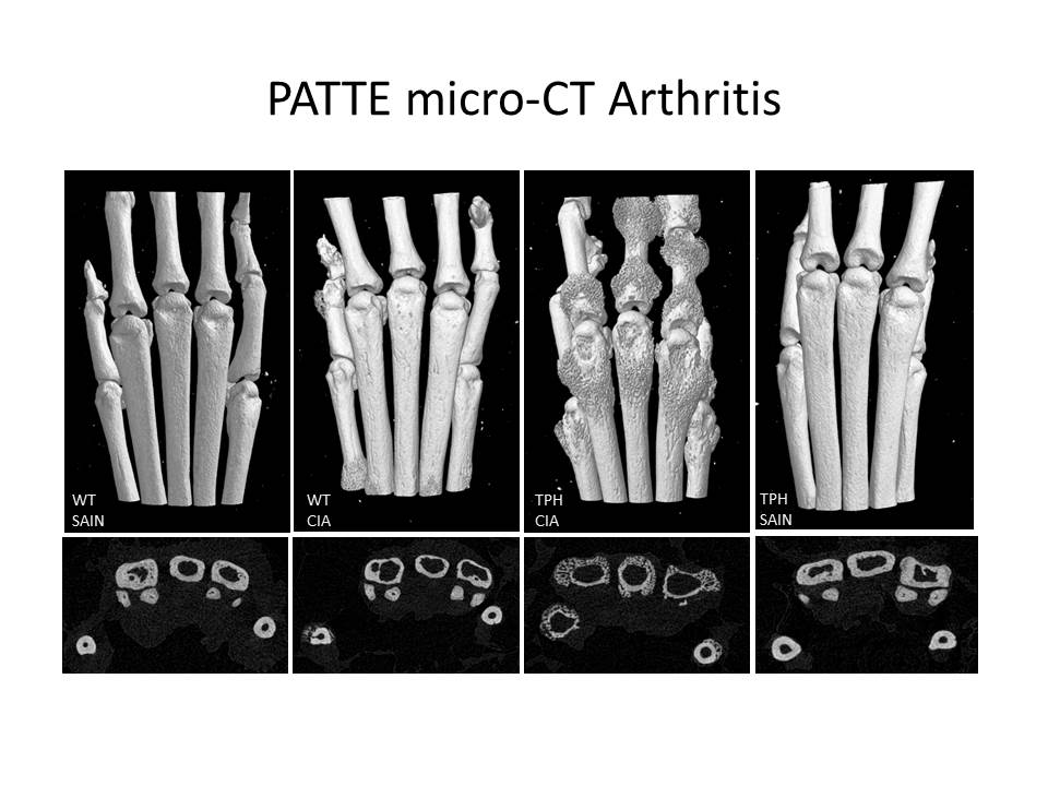
Arthritis and arthrosis study
We can measure bone density and eroded joints of lower limbs of mices with arthritis (inflammation) and/or mecanical arthrosis.
arthritis study : Paws of mices (without arthritis and with arthritis (=CIA))
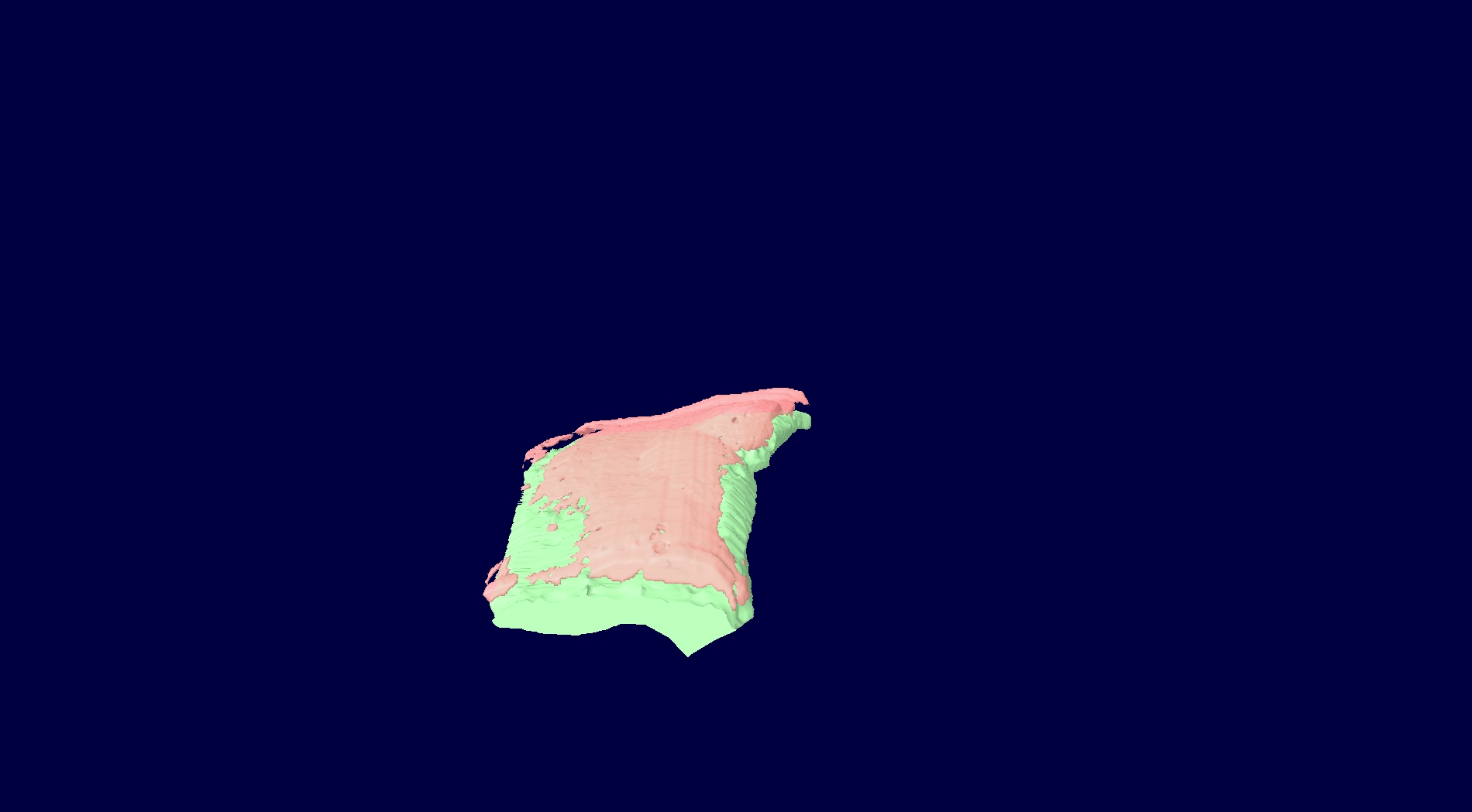
At cartilage knee level, we are setting up analysis methods of calcified and non-calcified areas.
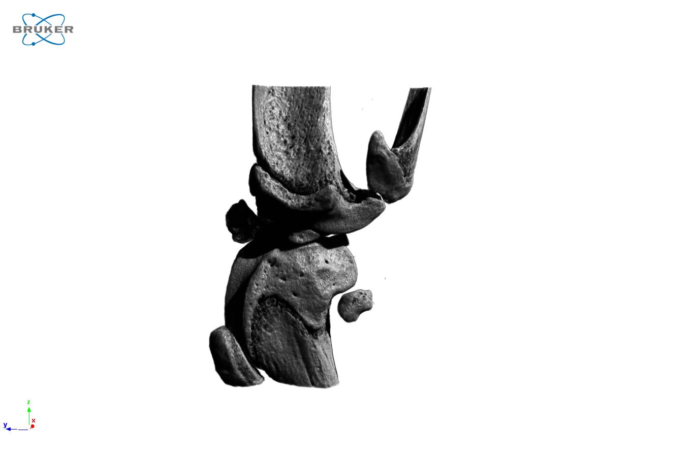
Knee of female hfe mouse
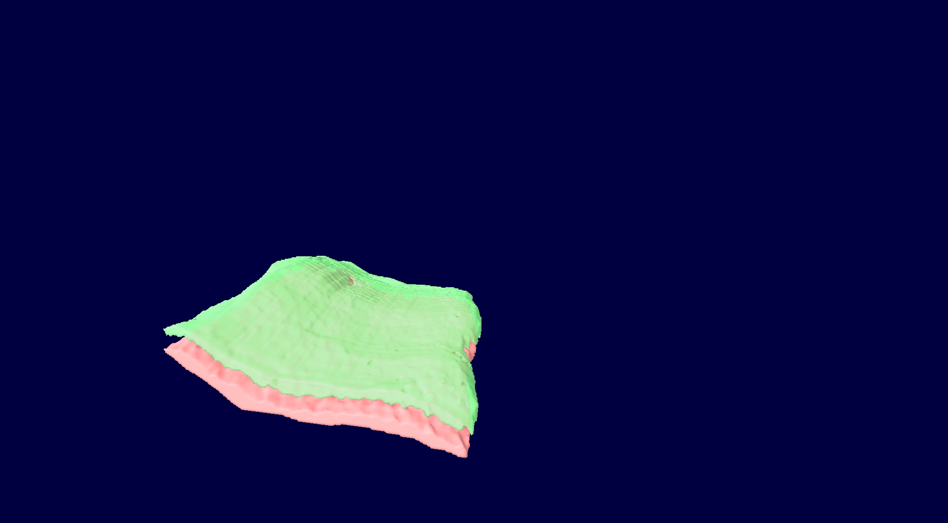
External plate
Internal plate
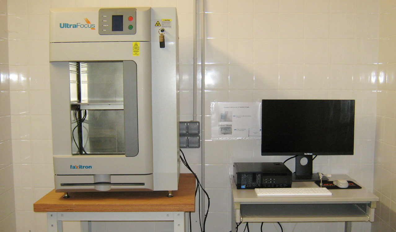
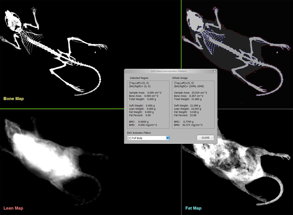
UltraFocus DXA FAXITRON
Descriptive : The UltraFocus DXA system (Faxitron) is used for ultra-high resolution imaging and dual-energy x-ray absorptiometry (DXA). It provides in vivo bone mineral density and body composition results from total body imaging (lean and fat mass) of little animal such as mice. It allows the researcher the opportunity to make multiple measurements in situ during the life of the animal, providing true longitudinal measures. As a digital radiography system, high quality images of animals can be made.